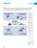
[en] Glioblastoma (GBM) is the most aggressive primary brain tumor characterized by infiltrative growth of malignant glioma cells into the surrounding brain parenchyma. In this study, our analysis of GBM patient cohorts revealed a significantly higher expression of Glycosyltransferase 8 domain containing 1 (GLT8D1) compared to normal brain tissue and could be associated with impaired patient survival. Increased in vitro expression of GLT8D1 significantly enhanced migration of two different sphere-forming GBM cell lines. By in silico analysis we predicted the 3D-structure as well as the active site residues of GLT8D1. The introduction of point mutations in the predicted active site reduced its glycosyltransferase activity in vitro and consequently impaired GBM tumor cell migration. Examination of GLT8D1 interaction partners by LC-MS/MS implied proteins associated with cytoskeleton and intracellular transport as potential substrates. In conclusion, we demonstrated that the enzymatic activity of glycosyltransferase GLT8D1 promotes GBM cell migration.
Disciplines :
Oncology
Author, co-author :
ILINA, Elena ; University of Luxembourg ; Department of Cancer Research (DoCR), Luxembourg Institute of Health (LIH), 1526 Luxembourg, Luxembourg. ; Luxembourg Centre of Neuropathology (LCNP), 1526 Luxembourg, Luxembourg.
CIALINI, Camille ; University of Luxembourg ; Department of Cancer Research (DoCR), Luxembourg Institute of Health (LIH), 1526 Luxembourg, Luxembourg. ; Luxembourg Centre of Neuropathology (LCNP), 1526 Luxembourg, Luxembourg.
GERLOFF, Dietlind ; University of Luxembourg > Luxembourg Centre for Systems Biomedicine > Bioinformatics Core ; Foundation for Applied Molecular Evolution (FfAME), Alachua, FL 32615, USA.
Duarte Garcia-Escudero, Maitane; Department of Cancer Research (DoCR), Luxembourg Institute of Health (LIH), 1526 Luxembourg, Luxembourg. ; Luxembourg Centre of Neuropathology (LCNP), 1526 Luxembourg, Luxembourg.
Jeanty, Céline; Quantitative Biology Unit, Luxembourg Institute of Health (LIH), 1526 Luxembourg, Luxembourg.
Thézénas, Marie-Laëtitia; Quantitative Biology Unit, Luxembourg Institute of Health (LIH), 1526 Luxembourg, Luxembourg.
Lesur, Antoine; Quantitative Biology Unit, Luxembourg Institute of Health (LIH), 1526 Luxembourg, Luxembourg.
Puard, Vincent; Quantitative Biology Unit, Luxembourg Institute of Health (LIH), 1526 Luxembourg, Luxembourg.
Bernardin, François; Quantitative Biology Unit, Luxembourg Institute of Health (LIH), 1526 Luxembourg, Luxembourg.
Moter, Alina; Department of Cancer Research (DoCR), Luxembourg Institute of Health (LIH), 1526 Luxembourg, Luxembourg. ; Luxembourg Centre of Neuropathology (LCNP), 1526 Luxembourg, Luxembourg.
Schuster, Anne; Department of Cancer Research (DoCR), Luxembourg Institute of Health (LIH), 1526 Luxembourg, Luxembourg. ; NORLUX Neuro-Oncology Laboratory, Department of Cancer Research (DoCR), Luxembourg Institute of Health (LIH), 1526 Luxembourg, Luxembourg.
Dieterle, Monika; Department of Cancer Research (DoCR), Luxembourg Institute of Health (LIH), 1526 Luxembourg, Luxembourg. ; NORLUX Neuro-Oncology Laboratory, Department of Cancer Research (DoCR), Luxembourg Institute of Health (LIH), 1526 Luxembourg, Luxembourg.
GOLEBIEWSKA, Anna ; University of Luxembourg ; Department of Cancer Research (DoCR), Luxembourg Institute of Health (LIH), 1526 Luxembourg, Luxembourg. ; NORLUX Neuro-Oncology Laboratory, Department of Cancer Research (DoCR), Luxembourg Institute of Health (LIH), 1526 Luxembourg, Luxembourg.
GERARDY, Jean-Jacques ; University of Luxembourg ; Luxembourg Centre of Neuropathology (LCNP), 1526 Luxembourg, Luxembourg. ; National Center of Pathology (NCP), Laboratoire National de Santé (LNS), 3555 Dudelange, Luxembourg.
DITTMAR, Gunnar ; University of Luxembourg ; Quantitative Biology Unit, Luxembourg Institute of Health (LIH), 1526 Luxembourg, Luxembourg.
NICLOU, Simone P. ; University of Luxembourg > Faculty of Science, Technology and Medicine (FSTM) > Department of Life Sciences and Medicine (DLSM) ; Department of Cancer Research (DoCR), Luxembourg Institute of Health (LIH), 1526 Luxembourg, Luxembourg. ; NORLUX Neuro-Oncology Laboratory, Department of Cancer Research (DoCR), Luxembourg Institute of Health (LIH), 1526 Luxembourg, Luxembourg.
Müller, Tanja; Department of Cancer Research (DoCR), Luxembourg Institute of Health (LIH), 1526 Luxembourg, Luxembourg. ; Luxembourg Centre of Neuropathology (LCNP), 1526 Luxembourg, Luxembourg.
MITTELBRONN, Michel ; University of Luxembourg > Luxembourg Centre for Systems Biomedicine (LCSB) > Neuropathology ; Department of Cancer Research (DoCR), Luxembourg Institute of Health (LIH), 1526 Luxembourg, Luxembourg. ; Luxembourg Centre of Neuropathology (LCNP), 1526 Luxembourg, Luxembourg. ; National Center of Pathology (NCP), Laboratoire National de Santé (LNS), 3555 Dudelange, Luxembourg.
Abdul Rahim, S.A., Dirkse, A., Oudin, A., Schuster, A., Bohler, J., Barthelemy, V., Müller, A., Vallar, L., Janji, B., Golebiewska, A., Niclou, S.P., Regulation of hypoxia-induced autophagy in glioblastoma involves ATG9A. Br. J. Cancer 117 (2017), 813–825, 10.1038/bjc.2017.263.
Auernheimer, V., Lautscham, L.A., Leidenberger, M., Friedrich, O., Kappes, B., Fabry, B., Goldmann, W.H., Vinculin phosphorylation at residues Y100 and Y1065 is required for cellular force transmission. J. Cell Sci. 128 (2015), 3435–3a443, 10.1242/jcs.172031.
Benes, P., Vetvicka, V., Fusek, M., Cathepsin D – many functions of one aspartic protease. Crit. Rev. Oncol. Hematol. 68 (2008), 12–28, 10.1016/j.critrevonc.2008.02.008.
Berman, H.M., Westbrook, J., Feng, Z., Gilliland, G., Bhat, T.N., Weissig, H., Shindyalov, I.N., Bourne, P.E., The protein Data Bank. Nucleic Acids Res. 28 (2000), 235–242, 10.1093/nar/28.1.235.
Bougnaud, S., Golebiewska, A., Oudin, A., Keunen, O., Harter, P.N., Mäder, L., Azuaje, F., Fritah, S., Stieber, D., Kaoma, T., et al. Molecular crosstalk between tumour and brain parenchyma instructs histopathological features in glioblastoma. Oncotarget 7 (2016), 31955–31971, 10.18632/oncotarget.7454.
Bowman, R.L., Wang, Q., Carro, A., Verhaak, R.G.W., Squatrito, M., GlioVis data portal for visualization and analysis of brain tumor expression datasets. Neuro Oncol. 19 (2017), 139–141, 10.1093/neuonc/now247.
Brenner, D., Weishaupt, J.H., Update on amyotropic lateral sclerosis genetics. Curr. Opin. Neurol. 32 (2019), 735–739, 10.1186/1750-1326-8-28.
Campos, B., Wan, F., Farhad, M., Ernst, A., Zeppernick, F., Tagscherer, K.E., Ahmadi, R., Lohr, J., Dictus, C., Gdynia, G., et al. Differentiation therapy exerts antitumor effects on stem-like glioma cells. Clin. Cancer Res. 16 (2010), 2715–2728, 10.1158/1078-0432.CCR-09-1800.
Cantarel, B.L., Coutinho, P.M., Rancurel, C., Bernard, T., Lombard, V., Henrissat, B., The Carbohydrate-Active EnZymes database (CAZy): an expert resource for glycogenomics. Nucleic Acids Res. 37 (2009), D233–D238, 10.1093/nar/gkn663.
Chaikuad, A., Froese, D.S., Berridge, G., von Delft, F., Oppermann, U., Yue, W.W., Conformational plasticity of glycogenin and its maltosaccharide substrate during glycogen biosynthesis. Proc. Natl. Acad. Sci. USA 108 (2011), 21028–21033, 10.1073/pnas.1113921108.
Cooper-Knock, J., Moll, T., Ramesh, T., Castelli, L., Beer, A., Robins, H., Fox, I., Niedermoser, I., van Damme, P., Moisse, M., et al. Mutations in the glycosyltransferse domain of GLT8D1 are associated with familial amyothrophic lateral sclerosis. Cell Rep. 26 (2019), 2298–2306, 10.1016/j.celrep.2019.02.006.
Cox, J., Mann, M., MaxQuant enables high peptide identification rates individualized p.p.b.-range mass accuracies and proteome-wide protein quantification. Nat. Biotechnol. 26 (2008), 1367–1372, 10.1038/nbt.1511.
Cox, J., Neuhauser, N., Michalski, A., Scheltema, R.A., Olsen, J.V., Mann, M., Andromeda: a peptide search engine integrated into the MaxQuant environment. J. Proteome Res. 10 (2011), 1794–1805, 10.1021/pr101065j.
Crowe, A.R., Yue, W., Semi-quantitative determination of protein expression UsingImmunohistochemistry staining and analysis: an integrated protocol. Bio Protoc., 9, 2019, e3465, 10.21769/BioProtoc.3465.
Dereeper, A., Guignon, V., Blanc, G., Audic, S., Buffet, S., Chevenet, F., Dufayard, J.F., Guindon, S., Lefort, V., Lescot, M., et al. Phylogeny.fr: robust phylogenetic analysis for the non-specialist. Nucleic Acids Res. 36 (2008), W465–W469, 10.1093/nar/gkn180.
Devraj, K., Poznanovic, S., Spahn, C., Schwall, G., Harter, P.N., Mittelbronn, M., Antoniello, K., Paganetti, P., Muhs, A., Heilemann, M., et al. BACE-1 is expressed in the blood–brain barrier endothelium and is upregulated in a murine model of Alzheimer's disease. J. Cereb. Blood Flow Metab. 36 (2016), 1281–1294, 10.1177/0271678X15606463.
Guindon, S., Dufayard, J.F., Lefort, V., Anisimova, M., Hordijk, W., Gascuel, O., New algorithms and methods to estimate maximum-likelihood phylogenies: assessing the performance of PhyML 3.0. Syst. Biol. 59 (2010), 307–321, 10.1093/sysbio/syq010.
Hu, H., Li, Z., Zhou, Y., Zhang, Y., Zhao, L., Zhao, W., Huang, Y., Song, X., GLT8D1 overexpression as a novel prognostic biomarker in human cutaneous melanoma. Melanoma Res. 29 (2019), 612–620, 10.1097/CMR.0000000000000631.
Hwang, S., Mahadevan, S., Qadir, F., Hutchison, I.L., Costea, D.E., Neppelberg, E., Liavaag, P.G., Waseem, A., The, M.T., Identification of FOXM1-induced epigenetic markers for head and neck squamous cell carcinomas. Cancer 119 (2013), 4249–4258, 10.1002/cncr.28354.
Larkin, M.A., Blackshields, G., Brown, N.P., Chenna, R., McGettigan, P.A., McWilliam, H., Valentin, F., Wallace, I.M., Wilm, A., Lopez, R., et al. Clustal W and clustal X version 2.0. Bioinformatics 23 (2007), 2947–2948, 10.1093/bioinformatics/btm404.
Lemjabbar-Alaoui, H., McKinney, A., Yang, Y.W., Tran, V.M., Phillips, J.J., Glycosylation alterations in lung and brain cancer. Adv. Cancer Res. 126 (2015), 305–344, 10.1016/bs.acr.2014.11.007.
Li, T., Guo, H., Song, Y., Zhao, X., Shi, Y., Hu, S., Nie, Y., Fan, D., Wu, K., Loss of vinculin and membrane-bound β-catenin promotes metastasis and predicts poor prognosis in colorectal cancer. Mol. Cancer, 13, 2014, 263, 10.1186/1476-4598-13-263.
Louis, D.N., Ohgaki, H., Wrestler, O.D., Cavenee, W.K., Ellison, D.W., Figarella-Branger, D., Perry, A., Reifenberger, G., von Deimling, A., Chapter 1: Diffuse astrocytic and oligodendrogial tumours. WHO Classification of Tumors of the Central Nervous System, 4th edn, 2016, International agency for research on cancer, Lyon, 16–77.
Moll, T., Shaw, P.J., Cooper-Knock, J., Disrupted glycosylation of lipids and proteins is a cause of neurodegeneration. Brain 143 (2019), 1332–1340, 10.1093/brain/awz358.
Okonechnikov, K., Golosova, O., Fursov, M., UGENE team. Unipro UGENE: a unified bioinformatics toolkit. Bioinformatics 28 (2012), 1166–1167, 10.1093/bioinformatics/bts091.
Pettersen, E.F., Goddard, T.D., Huang, C.C., Couch, G.S., Greenblatt, D.M., Meng, E.C., Ferrin, T.E., UCSF Chimera–a visualization system for exploratory research and analysis. J. Comput. Chem. 25 (2004), 1605–1612, 10.1002/jcc.20084.
Pinho, S., Reis, C., Glycosylation in cancer: mechanisms and clinical implications. Nat. Rev. Cancer 15 (2015), 540–555, 10.1038/nrc3982.
Pomaznoy, M., Ha, B., Peters, B., GOnet: a tool for interactive Gene Ontology analysis. BMC Bioinformatics, 19, 2018, 470, 10.1186/s12859-018-2533-3.
Ren, J., Wen, L., Gao, X., Jin, C., Xue, Y., Yao, X., DOG 1.0: illustrator of protein domain structures. Cell Res. 19 (2009), 271–273, 10.1038/cr.2009.6.
Rowe, L., Burkhart, G., Analyzing protein glycosylation using UHPLC: a review. Bioanalysis 10 (2018), 1691–1703, 10.4155/bio-2018-0156.
Rubashkin, M.G., Cassereau, L., Bainer, R., DuFort, C.C., Yui, Y., u, G., Paszek, M.J., Davidson, M.W., Chen, Y.Y., Weaver, V.M., Force engages vinculin and promotes tumor progression by enhancing PI3-kinase activation of phosphatidylinositol (3, 4, 5)-triphosphate. Cancer Res. 74 (2015), 4597–4611, 10.1158/0008-5472.CAN-13-3698.
Sanzey, M., Abdul Rahim, S.A., Oudin, A., Dirkse, A., Kaoma, T., Vallar, L., Herold-Mende, C., Bjerkvig, R., Golebiewska, A., Niclou, S.P., Comprehensive analysis of glycolytic enzymes as therapeutic targets in the treatment of glioblastoma. PLoS One, 10, 2015, e0123544, 10.1371/journal.pone.0123544.
Sasayama, D., Hori, H., Yamamoto, N., Nakamura, S., Teraishi, T., Tatsumi, M., Hattori, K., Ota, M., Higuchi, T., Kunugi, H., ITIH3 polymorphism may confer susceptibility to psychiatric disorders by altering the expression levels of GLT8D1. J. Psychiatr. Res. 50 (2014), 79–83, 10.1016/j.jpsychires.2013.12.002.
Schneider, C., Rasband, W., Eliceiri, K., NIH Image to ImageJ: 25 years of image analysis. Nat. Methods 9 (2012), 671–675, 10.1038/nmeth.2089.
Sethi, M.K., Buettner, F.F.R., Krylov, V.B., Takeuchi, H., Nifantiev, N.E., Haltiwanger, R.S., Gerardy-Schahn, R., Bakker, H., Identification of glycosyltransferase 8 family members as xylosyltransferases acting on O-glycosylated notch epidermal growth factor repeats. J. Biol. Chem. 285 (2010), 1582–1586, 10.1074/jbc.C109.065409.
Seznec, J., Silkenstedt, B., Naumann, U., Therapeutic effects of the Sp1 inhibitor mithramycin A in glioblastoma. J. Neurooncol. 101 (2011), 365–377, 10.1007/s11060-010-0266-x.
Singh, J.P., Zhang, K., Wu, J., Yang, X., O-GlcNAc signaling in cancer metabolism and epigenetics. Cancer Lett. 356:2 Pt A (2015), 244–250, 10.1016/j.canlet.2014.04.014.
Stelzer, G., Rosen, R., Plaschkes, I., Zimmerman, S., Twik, M., Fishilevich, S., Iny Stein, T., Nudel, R., Lieder, I., Mazor, Y., et al. The GeneCards suite: from gene data mining to disease Genome sequence analysis. Curr. Protoc. Bioinformatics 54 (2016), 1.30.1–1.30.33, 10.1002/cpbi.5.
Taujale, R., Venkat, A., Huang, L.-C., Zhou, Z., Yeung, W., Rasheed, K.M., Li, S., Edison, A.S., Moremen, K.W., Kannan, N., Deep evolutionary analysis reveals the design principles of fold A glycosyltransferases. Elife, 9, 2020, e54532, 10.7554/eLife.54532.
Vajaria, B.N., Patel, K.A., Patel, P.S., Role of aberrant glycosylation enzymes in oral cancer progression. J. Cancirog., 17, 2018, 5, 10.4103/jcar.JCar_7_18.
Vollmann-Zwerenz, A., Leidgens, V., Feliciello, G., Klein, C.A., Hau, P., Tumor cell invasion in glioblastoma. Int. J. Mol. Sci., 21, 2020, E1932, 10.3390/ijms21061932.
Waterhouse, A.M., Procter, J.B., Martin, D.M.A., Clamp, M., Barton, G.J., Jalview Version 2 – a multiple sequence alignment editor and analysis workbench. Bioinformatics 25 (2009), 1189–1191, 10.1093/bioinformatics/btp033.
Waterhouse, A., Bertoni, M., Bienert, S., Studer, G., Tauriello, G., Gumienny, R., Heer, F.T., de Beer, T.A.P., Rempfer, C., Bordoli, L., et al. SWISS-MODEL: homology modelling of protein structures and complexes. Nucleic Acids Res. 46 (2018), W296–W303, 10.1093/nar/gky427.
Yang, C.P., Li, X., Wu, Y., Shen, Q., Zeng, Y., Xiong, Q., Wei, M., Chen, C., Liu, J., Huo, Y., et al. Comprehensive integrative analyses identify GLT8D1 and CSNK2B as schizophrenia risk genes. Nat. Commun., 9, 2018, 838, 10.1038/s41467-018-03247-3.
Yu, H., Takeuchi, M., LeBarron, J., Kantharia, J., London, E., Bakker, H., Haltiwanger, R.S., Li, H., Takeuchi, H., Notch-modifying xylosyltransferase-substrate complexes support an SNi-like retaining mechanism. Nat. Chem. Biol. 11 (2015), 847–854, 10.1038/nchembio.1927.
Zahn-Zabal, M., Michel, P.A., Gateau, A., Nikitin, F., Schaeffer, M., Audot, E., Gaudet, P., Duek Roggli, P., Teixeira, D., Rech de Laval, V., et al. The neXtProt knowledgebase in 2020: data, tools and usability improvements. Nucleic Acids Res. 48 (2020), D328–D334, 10.1093/nar/gkz995.
Zhang, Z., Izaguirre, G., Lin, S.-Y., Lee, H.Y., Schaefer, E., Haimovich, B., The phosphorylation of vinculin on Tyrosine residues 100 and 1065, mediated by SRC kinase, affects cell spreading. Mol. Biol. Cell 15 (2004), 4234–4247, 10.1091/mbc.e04-03-0264.
Zimmermann, L., Stephens, A., Nam, S.Z., Rau, D., Kübler, J., Lozajic, M., Gabler, F., Söding, J., Lupas, A.N., Alva, V., A completely reimplemented MPI bioinformatics toolkit with a new HHpred server at its core. J. Mol. Biol. 430 (2018), 2237–2243, 10.1016/j.jmb.2017.12.007.